Keywords: nevi of Ota, nevus of Ota treatment, dermal melanocytosis, Q-switched laser therapy
What Is Nevi of Ota?
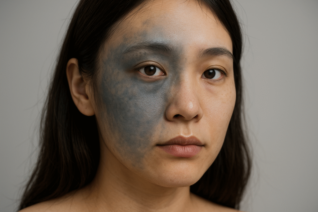
Nevi of Ota, also known as oculodermal melanocytosis, is a rare form of dermal melanocytosis characterized by bluish-gray hyperpigmentation affecting the skin innervated by the ophthalmic and maxillary branches of the trigeminal nerve. The condition primarily involves the periorbital area, temples, forehead, and sometimes the sclera and mucous membranes.
Etiology and Pathogenesis
The pathophysiology of nevi of Ota lies in the aberrant presence of melanocytes in the upper dermis. While the exact mechanism remains unclear, it is generally believed to result from the incomplete migration of melanocytes from the neural crest during embryogenesis. Although most cases are congenital, some patients may exhibit delayed onset during adolescence or early adulthood, potentially triggered by hormonal changes, UV exposure, or inflammation.
Epidemiology and Clinical Presentation
Nevi of Ota is most commonly observed in individuals of Asian or African descent, with a marked female predominance. Clinically, it manifests as mottled or confluent patches of bluish-gray discoloration on one side of the face. In approximately 10% of cases, the pigmentation may appear bilaterally. The involvement of ocular structures—such as the sclera, conjunctiva, and uveal tract—necessitates ophthalmologic evaluation to rule out complications like glaucoma.
Diagnostic Criteria
Diagnosis is primarily clinical, supported by patient history and characteristic pigmentation patterns. Additional tools that may aid in evaluation include:
- Dermoscopy: To assess the pigment depth and exclude malignancies.
- Histopathology: Reveals spindle-shaped melanocytes within the dermis.
- Imaging: Rarely needed unless ocular or neurological involvement is suspected.
Nevus of Ota Treatment: Current Gold Standards
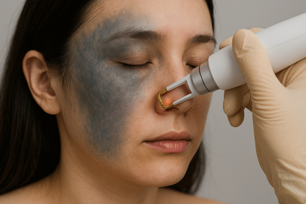
Q-Switched Laser Therapy
The mainstay of nevus of Ota treatment is pigment-targeted laser therapy, particularly Q-switched lasers, which deliver high-intensity pulses in nanoseconds to selectively fragment melanin granules. Options include:
- Q-switched Nd:YAG laser (1064 nm) – Suitable for darker skin types (Fitzpatrick III–VI).
- Q-switched Ruby laser (694 nm) – Effective for lighter skin types but carries a higher risk of post-inflammatory pigmentary changes.
- Q-switched Alexandrite laser (755 nm) – Balances efficacy and safety in medium skin tones.
Multiple sessions spaced 6–8 weeks apart are typically required for optimal results, with gradual lightening observed over months.
Fractional and Picosecond Lasers
Recent advancements include picosecond lasers, which may reduce treatment sessions and improve pigment clearance with less risk of adverse effects. Fractional modalities are also being explored for refractory cases.
Adjunctive and Supportive Therapies
While laser remains the gold standard, supportive measures can enhance outcomes and reduce recurrence:
- Broad-spectrum sunscreen: Prevents UV-induced pigment darkening and recurrence.
- Topical depigmenting agents: Hydroquinone, arbutin, or azelaic acid may offer limited adjunctive benefits.
- Camouflage techniques: High-pigment cosmetic coverage for patients ineligible for laser treatment.
Prognosis and Follow-up
Nevi of Ota is a benign condition with excellent outcomes when appropriately treated. However, recurrence or incomplete pigment clearance is possible. Regular follow-up is essential, especially in cases with ocular involvement, where intraocular pressure monitoring is advised to exclude glaucoma.
Summary
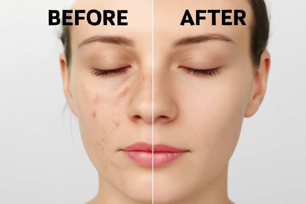
Nevi of Ota, though benign, can have a significant psychosocial impact due to its visible nature. Advances in nevus of Ota treatment, particularly laser-based modalities, have revolutionized clinical outcomes. Early diagnosis and personalized treatment planning are key to achieving optimal therapeutic results with minimal risk.
Frequently Asked Questions (FAQs)
Q1: Is nevi of Ota dangerous?
No, it is a benign pigmentary disorder. However, ocular involvement may require medical monitoring.
Q2: How many laser sessions are needed?
Typically 5–10 sessions are needed, depending on skin type, pigmentation depth, and response.
Q3: Is nevus of Ota permanent?
Without treatment, yes. Laser therapy can significantly reduce or eliminate pigmentation.